A multi-modality gene therapy study was conducted to determine whether there was a correlation between carrier distribution and gene transduction. To answer this question, we compared the bio-distribution and PK of a radiolabeled AAV to its subsequent iRFP gene transduction and expression in Sprague Dawley rats.
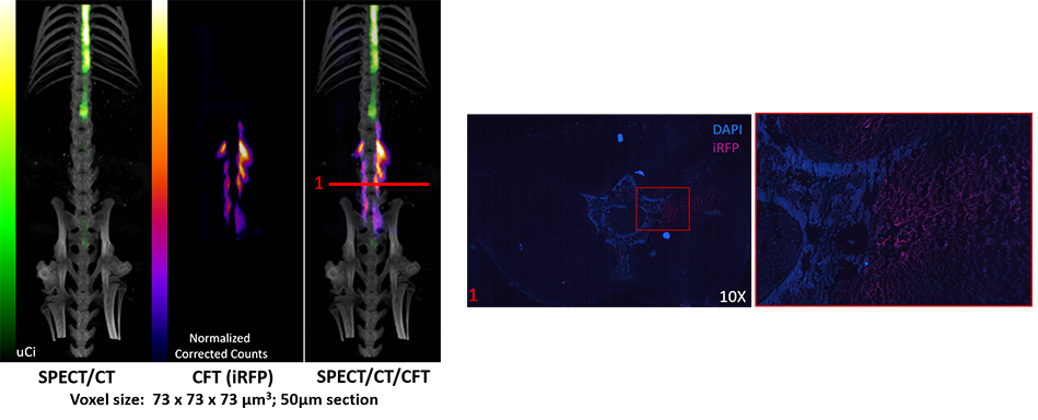
Subjects were intrathecally (IT) administered I125-AAV and AAViRFP together for in vivo imaging. SPECT and CT were used to observe the in vivo biodistribution of I125AAVs within hours of administration. Four weeks later, Cryofluorescence Tomography (CFT) and fluorescence microscopy were used to examine the transduction of AAVs and the subsequent expression of iRFP following euthanasia (Figure 1).
SPECT, CT and CFT data were co-registered to provide a 3D-visualization of biodistribution and transduction of I125-AAV and AAViRFP, respectively. SPECT/CT results showed that AAV was distributed throughout the spine, while CFT showed iRFP expression in the surrounding muscle tissue around the lumbar spinal region. This iRFP expression was further confirmed by section collection and microscopy imaging.
Ultimately, our multi-modal approach successfully demonstrated the ability to answer a complex question using a multitude of data including temporal, spatial, 2D, 3D, and high- and low-resolution images from each subject.
Figure 1