Oncology
Overview
Bringing new cancer therapies to the market is a complex and challenging process that requires a partner that can support you every step of the way. Invicro combines operational excellence with extensive scientific and medical expertise to support you with preclinical work through late phase clinical trials. Our consultative approach allows us to design, execute and deliver results that can be used to make critical decisions across all stages of the cancer drug development continuum. We have experience working with a wide-range of therapeutics, including small molecules, peptides, antibodies, multi-specific proteins, CAR-T cells and many more. Our comprehensive oncology biomarker services, include:
Radiopharmaceutical Therapy & Theranostics
Overview
Radiopharmaceutical therapies represent an effective way to treat solid cancers by using tumor targeting small molecules, peptides or biologics to deliver a cytotoxic payload that induces DNA damage in tumor cells, while limiting damage to normal and healthy tissue.
Streamlined development, evaluation and clinical translation of radiopharmaceuticals requires a partner that has extensive scientific, regulatory and operational expertise. Working with a CRO that provides a complete solution ensures data is delivered in a timely manner to make a go/no-go decision on your imaging or
therapeutic candidates.
Services Include:
- Radiochemistry development – conjugation, radiolabeling, stability and immunoreactivity
- Preclinical proof-of-concept studies – biodistribution, efficacy, toxicology
- Investigational New Drug (IND)/Clinical Trial Application (CTA) submission support with clinical protocol development
- Single-site, first-in-human through multi-site, late phase clinical study support
- Good manufacturing practice (GMP) radiochemistry development and production support for imaging agents
- Preclinical and clinical dosimetry and advanced quantitative image analysis
Watch Theranostics Panel Discussion
Info Sheet – Radiopharmaceutical Therapy
Imaging/Theranostic Agent Development Process

Capabilities
Radiochemistry
Our team has experience with novel and commercially available radioisotopes used for targeted radiotherapy. Our capabilities for novel radiotracers span preclinical to first-in-human clinical studies and GMP production with Invicro’s CMO partners. Invicro has experience working with the following isotopes:
- 225Ac
- 177Lu
- 131I
- 111ln
- 68Ga/177Lu
- 111In/225Ac
- 203Pb/212Pb
- 67Cu/64Cu
Preclinical Solutions
Bringing new therapeutic radiopharmaceuticals to the market is a complex and challenging process. When filing an Investigational New Drug (IND) or Clinical Trial Application (CTA) prior to the initiation of human trials, it is crucial to have strong non-clinical data to support a new candidate. Invicro provides complete preclinical support of your project, including:
-
- Study design and consultation
- Radiolabeling/conjugation to support alpha, beta, and gamma emitting isotopes
- in vitro/ex vivo cell and tissue-based assays
- Primary pharmacology studies to demonstrate MOA
- in vivo Imaging model development to support theranostic imaging
- Advanced image analytics
- Dosimetry
- IND support
- Development of chemistry, manufacturing and controls package for imaging agents
Early/Late Phase Clinical
Seamless transition of your radiopharmaceutical into the clinic requires an imaging partner with experience supporting first-in-human, single-site and late phase, multi-site global clinical studies. Invicro has the unique blend of scientific expertise and operational experience to deliver on studies from Phase 0-IV. Clinical radiopharmaceutical therapeutic development services include:
- First-in-human, single-site clinical trial support
- Study design and consultation
- Criteria-based centralized independent reviews and internal analysis
- Late stage, global multi-center clinical trial support with response criteria reads
- Advanced image analytics including dosimetry
- Safety profile and therapeutic efficacy studies
Dosimetry and Advanced Analytics
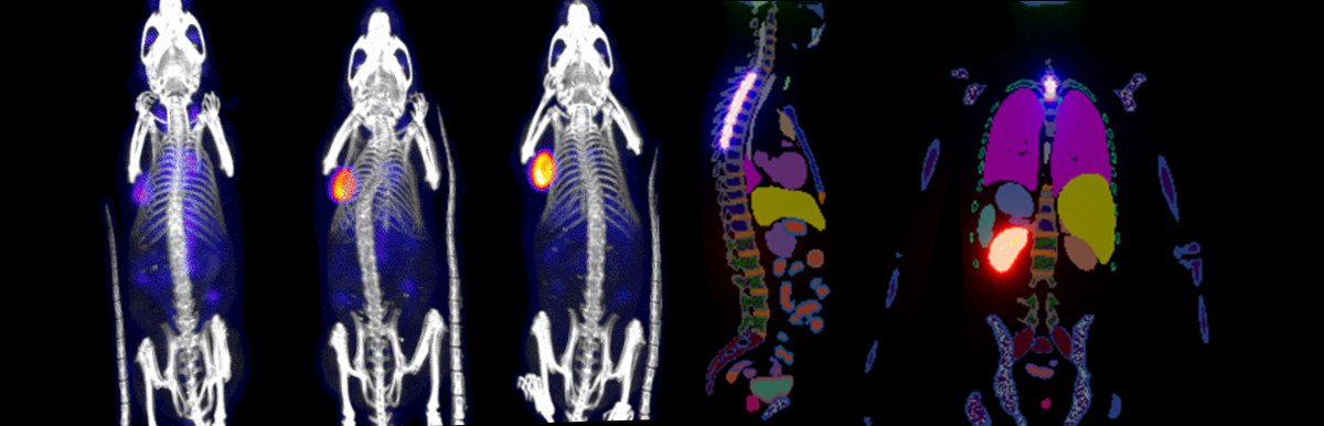
Our in-house image analytics team has experience supporting preclinical and clinical radiopharmaceutical therapy studies, spanning from customized and automated image processing techniques using deep learning to dosimetry analysis for safety profile analysis. Some advanced image analytic techniques include:
- Algorithms for automated and semi automated lesion identification
- Image quality control and co-registration
- Preclinical dosimetry safety studies and validated clinical workflows
- Extrapolating dosimetry of therapeutics from imaging agents
- Expertise across various species, radionuclides and novel administration routes
PSMA-PET Imaging Solutions
Invicro is an industry-leading CRO providing full-service PSMA-PET solutions from radiochemistry development to preclinical and Phase 0-IV clinical trials. We support researchers that are developing PSMA-targeted prostate cancer therapeutics or multi-specifics, and groups using PSMA expression as a biomarker for patient selection, and treatment response in prostate cancer clinical trials. Invicro advances PSMA-PET standard of care by combining advanced image analytics and artificial intelligence with standard PET SUV readouts to ensure you are receiving the most meaningful data from your images. In recent years, Invicro has overseen more clinical PSMA-PET scans (>3,600) than most other imaging CROs and is one of the only organizations to support an FDA submission combining AI and quantitative PET.
Our full-service PSMA-PET solutions include:
-
- Complete global core lab support for Phase 0-IV clinical trials
- Radiology expertise for study design, image analysis and sub-specialty reads (PCWG3)
- Advanced analytical tools for whole body image quantification to estimate disease burden, lesion localization, and response to therapy.
- Radiochemistry development and expert guidance on PSMA ligand access and utilization, including external manufacturing support
- Preclinical solutions for discovery research
- Extensive experience with Regulatory filing support (e.g., IND-enabling support)
Clinical Capabilities
Invicro’s clinical team has expertise in medical imaging and a vast range of tools to support your cancer drug discovery and development needs. With over 200 scientists and 3,500+ qualified imaging centers and clinical sites across the globe, Invicro has the advanced scientific, medical and regulatory expertise and project management scale to deliver studies from Phase 0-IV. Our technology platforms support key decision-making in:
- First-in-human, single-site clinical trial support
- Late stage, global multi-center clinical trial support with response criteria reads
- Advanced image analytics including dosimetry
- Safety profile and therapeutic efficacy studies
- Study design and consultation
- Criteria-based centralized independent reviews and internal analysis
Our imaging core lab offers criteria-driven reads for standard-of-care projects and our state-of-the-art in-house software enables 3D region-of-interest generation to support quantitative analysis methods, such as PERCIST. We also partner with subspecialist independent readers that focus on specific areas of expertise. Some of the validated criteria examples include:
- RECIST 1.0/1.1
- PCWG 2.0/3.0
- RANO
- LUGANO
- IMWG
- CHESON
- iRECIST
- irRECIST
- LYRIC
- irRC
Quantitative Oncology Image Analytics
Invicro’s medical image analysis team supports standard analysis of FDG PET imaging, including PERCIST and other SUV-based metrics, metabolic tumor volume and total lesion glycolysis. The image analysis team also specializes in developing and applying a broad range of tools to support advanced quantitative oncology tracer characterization and analytics across a broad range of modalities and studies. Some examples include:
- Powerful, multi-modal tumor and organ Segmentation tools
- A range of flexible Biomathematical Modeling tools
- Dosimetry calculations across radionuclide, administration route, and species
- Integrated Radiomics pipelines using standard and custom feature vectors
- Preclinical and clinical support for Tracer Characterization
- Sophisticated batch Image Triage and preprocessing methods
For more information, check out some of our most popular case studies. A full list is also available in our case studies section.
Discovery and Preclinical Capabilities
Invicro’s discovery and preclinical services team provides study design, study execution and data analysis services for a broad-range of solid and non-solid tumor studies. Our preclinical oncology team uses state-of-the-art imaging techniques to characterize the behavior of novel therapies, including antibodies, radioligand therapies, peptides, cell therapies (e.g., CAR-T), exosomes and other tumor-targeting modalities in diverse tumor model types (e.g., subcutaneous, orthotopic, metastatic). Non-clinical work supported by Invicro can be used to move into first-in-human studies to late-stage clinical multicenter trials. Our core capabilities include the measurement of multiple readouts, including:
- Biodistribution of test articles
- Target density/engagement
- Pharmacokinetics (PK)
- Pharmacodynamics (PD)
- Tumor growth properties
- Therapeutic efficacy
- Dosimetry
Advanced Pathology Services
Invicro provides comprehensive pathology services to support all phases of oncology and immuno-oncology drug development applications. Our multidisciplinary team of scientific and medical experts provide a unique approach by leveraging cutting-edge and proprietary molecular pathology detection and analysis technologies. From experimental design to developing and validating biomarker assays, we deliver actionable insights in both a research and CAP-CLIA environment. Our capabilities include:
- Core histology
- Specialty stains
- Multiplex Immunohistochemistry (IHC)
- Multiplex Immunofluorescence (IF)
- RNAScope® ISH
- BaseScope™ ISH
- CAP/CLIA assay development and validation
- Quanticell™ High sensitivity Immunohistochemistry (IHC)
- Pathologist interpretation
- Whole Slide Imaging (WSI)
- Image analysis
- Companion Diagnostic (CDx) development
- Digital biomarker development
Radiochemistry
Invicro’s radiochemistry team has experience with most commercially available radioisotopes used for PET/SPECT imaging and several used in targeted radiotherapy. Our capabilities span from first-in-human clinical studies to GMP implementation of novel radiotracers, including:
- A Microdose PET Study of the Safety, Immunogenicity, Biodistribution, and Radiation Dosimetry of 18F-FB-A20FMDV2 for Imaging the Integrin αvβ
- Clinical Translation of a Click-Labeled 18F-Octreotate Radioligand for Imaging Neuroendocrine Tumors
- 18F-ICMT-11, a Caspase-3–Specific PET Tracer for Apoptosis: Biodistribution and Radiation Dosimetry
- Biodistribution and radiation dosimetry of deuterium-substituted 18F-fluoromethyl-[1, 2-2H4]choline in healthy volunteer
Thought Leadership Content
Webinars
The Resurgence of Theranostics: Challenges and Successes of Clinical Application
Translating Radiolabeled Biologics – Strategies for Successful IND Submission
EXPLORER as a Transformational Imaging Technology for Drug Development
Segmentation of Lesions in Whole-Body PET/CT Using Deep Learning
Utilizing Imaging Agent Biomarkers for Improved Patient Management – Challenges and Successes
PSMA: Imaging Biomarker Development
The Changing Landscape of Response Assessment in Oncology
Imaging Biomarkers in Metastatic Prostate Cancer
Case Studies
Radiolabeling of a DOTA Conjugate Antibody for Theranostic Applications
Preclinical development and first-in-human study of 68Ga-MLN6907
Antigen Density Estimation – Clinical PET/CT Protocol Design for a First-In-Human Diagnostic Peptide
Infographics
Radiopharmaceutical Drug Development
Complete Radiopharmaceutical Therapy Services