Multi-modality imaging examines the anatomical, functional and molecular features of the targeted disease and it is achieved by executing different imaging modalities. There are typically two modalities used for this method: (1) anatomical imaging that utilizes CT or MRI and (2) molecular/structural imaging that utilizes PET, SPECT, fMRI and more. The two modalities are co-registered to verify alignment and the ROIs are defined by using the information of both imaging techniques. Afterward, the signal is quantified using molecular/structural imaging.
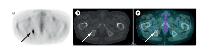
For example, multi-modality imaging was used to determine metastasis on a patient with non-small-cell lung cancer. In figure 1. below the FDG-PET (image a) and CT (image b) were fused to provide functional and anatomical information. The fusion (image c) showed a metastasis was found on the right ischial tuberosity. Merging the strength of the modalities has provided diagnostic precision in many studies and aids in tumor classification and treatment plan development.
Click Here for more information
Figure 1