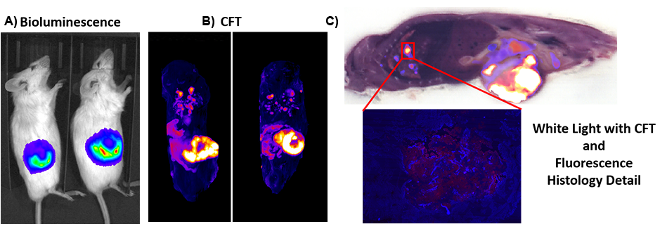
A tumor cell line (4T1 mouse mammary tumor) expressing luciferase, an oxidative enzyme that produces bioluminescence and DsRed, a fluorescent protein, were used to compare the sensitivity and resolution of bioluminescence imaging (BLI) and Cryofluorescence Tomography (CFT) (Figure 1). Half a million cells were injected into immunodeficient NSG mice via their hearts’ left ventricles. A) The tumor metastasis was monitored regularly using a bioluminescence imaging system for 10 days. B) The animals were then euthanized, frozen and embedded in optimum cutting temperature (OCT) compound for CFT.
C) Sequential sectioning (50 μm) and image acquisition (white light and fluorescence) were captured. The white light and fluorescence images were then processed and reconstructed into a 3D volume. While bioluminescence showed high signal to noise (S/N), the imaging system was only able to detect and image the main tumor that is close to the surface; lacking the resolution to resolve metastases below 1 mm from the surface. On the other hand, the CFT results provided higher resolution imaging of the main tumor and demonstrated the ability to detect and localize tumor metastasis as small as 300 μm in the lungs. The corresponding white light images provided anatomical reference to better resolve lesion location.
Figure 1