Antisense oligonucleotides (ASOs) are utilized for treating central nervous system (CNS) disorders due to their specificity and long-lasting pharmacological effects. Due to the size and charge of ASO particles, intrathecally (IT) administration is used to bypass the blood-brain-barrier (BBB). The anatomical and functional properties of the intrathecal space make it difficult to determine the pharmacokinetics (PK) and pharmacodynamics (PD) of ASOs. Therefore, the goal of this study was to determine the PK and PD of ASOs using a combination of in vivo and ex vivo imaging techniques.
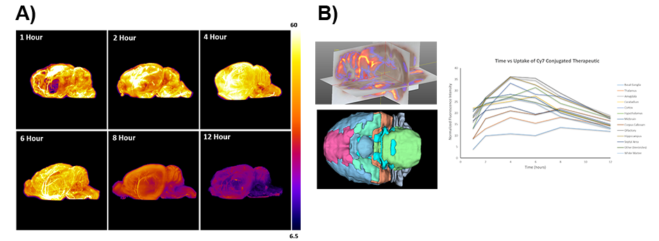
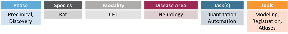
Two different ASOs, that target the Malat1 non-coding RNA and GABA-A receptor subunit Gabra1 mRNA, were used to demonstrate the PK and PD effects of ASOs. The imaging of 125I-Malat1-ASO using SPECT/CT demonstrated time and dose dependent localization in the neuroaxis and cortical structures. To provide higher 3D resolution, Cryofluorescence Tomography (CFT) was conducted in rats that were IT-administered with Gabra-1 ASO labeled with Cy7. Rats were euthanized at 1, 2, 4, 8, and 12 hours following test article injection. Sequential coronal sectioning (25 μm) and image acquisition (white light and fluorescence) were conducted throughout the rat brain (Figure 1A). Following image acquisition, all the acquired 2D images were processed, reconstructed into 3D volumes and presented as maximum intensity projections (MIP) (Figure 1B). Results demonstrate the uptake and clearance of ASOs throughout the brain. A region-based analysis was conducted by co-registering the 3D volume to a rat brain atlas. Multiplanar reconstruction (MPR) images were also created to provide a better visualization of ASO localization in the brain.
Figure 1