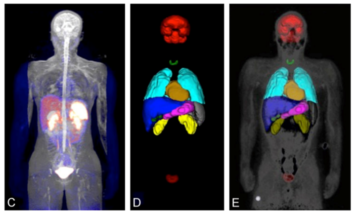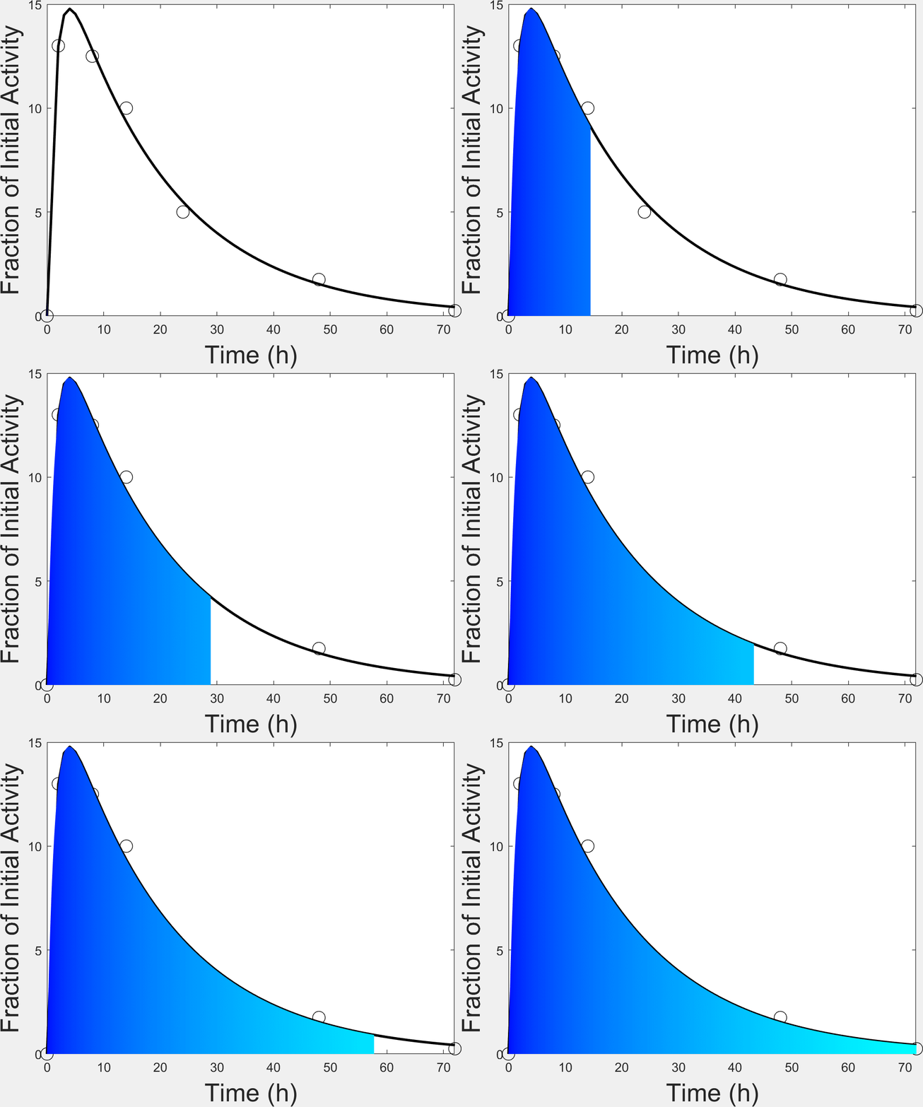
Dosimetry analysis is a multi-step process that utilizes different tools to generate estimates of tissue time-activity curves and the associated radiation absorbed dose. This process can be broken down into three steps: (1) calculation of mean residence time (MRT) values, (2) production of dosimetry output using OLINDA, and (3) quality control of dosimetry output by image processing specialists.
MRT values are detected in the source organ or tissue of interest of all subjects. If the subjects are not human, then the MRT values equivalent to human are found. Images with regions of interest (ROIs) are used to generate time-activity curves that are not decay corrected. The ROIs are specific to the type of study being conducted. Once all the needed MRT values are calculated, the information is entered in OLINDA to produce effective human doses. During this process the appropriate nuclide, body phantom and kinetic data are selected. The input data is verified to make sure it correlates with the images used and that the measurements do not exceed its isotope’s decay. This method supports the biodistribution of radioactive isotopes over time following clinical administration, specifically for radiation therapy.
Click here for more information
Analysis / Segmentation

Computation of MRT

68Ga-DOTATOC Dosimetry, Esfehani et al., 2017
