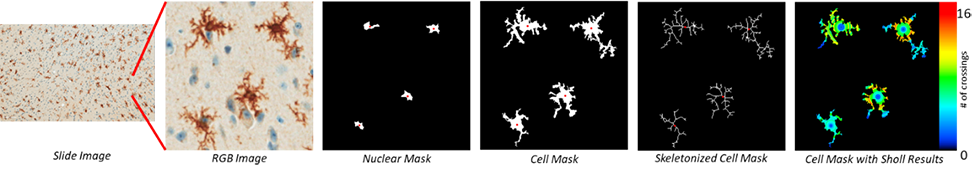
Analysis of microglia morphology was adapted into an automated workflow. Images of stained microglia were processed to identify nuclei and isolate cell structures that enabled the generation of nuclear masks, cell masks and skeletonized cell masks. Metrics such as area, maximum distance and number of branch points were extrapolated from the masks to perform Linear Scholl analysis (Figure 1). Results were overlaid over the cell mask to display the number of dendritic crossings (Figure 2).
Figure 1
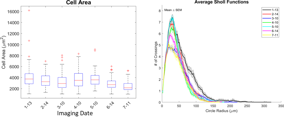
Figure 2