A fully automated tooth segmentation tool was developed to aid in the detection and segmentation of rhesus monkey teeth in CT images. The tool utilized multi-atlas segmentation at several resolutions and levels of zoom to minimize the processing time while still maintaining high accuracy in the segmentations.
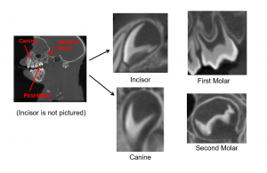
Twelve CT images were provided with regions of interest manually defined to act as a reference library. The four teeth segmented were the left incisor, the left canine and two molars on the left side. The multi-atlas segmentation process is as follows:
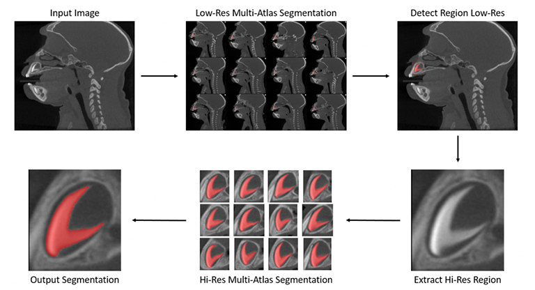
- Register each reference data set to the input image using an affine transform followed by a deformable transform. Apply the same transforms to the region of interest data.
- Average several the transformed reference ROIs together to construct a probability map on the input image of each region of interest.
- Threshold the probability map to build a deterministic output region of interest.
Multi-Atlas segmentation was run on the entire field of view at a low resolution for all 4 regions. A bounding box for each output region was determined and the region was extracted. Multi-Atlas segmentation was then run again on each focused high-resolution region to determine the final segmentation.
Example 1

Example 2

Figure 1

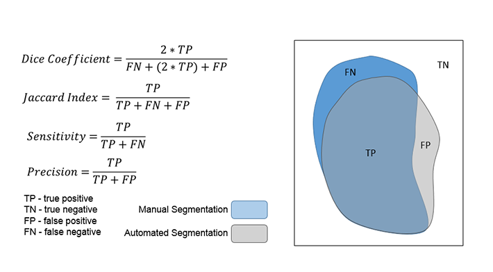
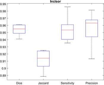
Performance was evaluated by running each member of the reference library through the tool while excluding the input from the reference library. The output automated segmentations were compared to the provided manual segmentations at a voxel-by-voxel level and metrics such as the Dice coefficient, Jaccard index, sensitivity and precision were computed.
Example 3

